Файл:Proteaosome 1fnt side.png
Перейти к навигации
Перейти к поиску
Размер этого предпросмотра: 250 × 600 пкс. Другие разрешения: 100 × 240 пкс | 500 × 1200 пкс.
Исходный файл (500 × 1200 пкс, размер файла: 724 КБ, MIME-тип: image/png)
Краткое описание
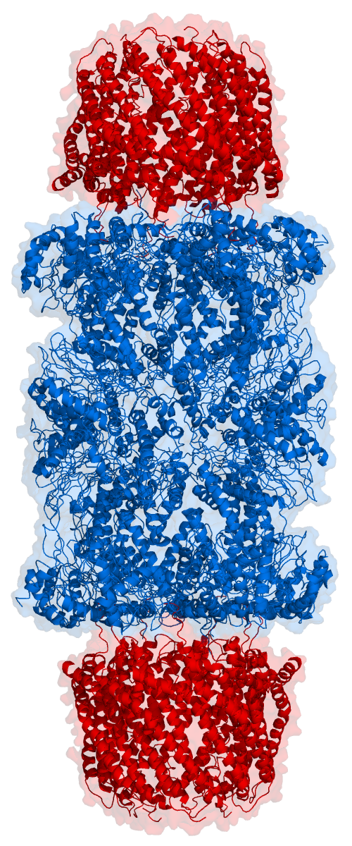
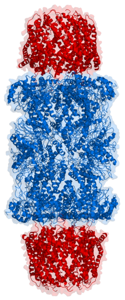
| Описание | Cartoon representation of a proteasome. This cylinder-shaped protein has its active sites sheltered inside the tube (blue). The caps (red) on the ends regulate entry into the destructive chamber, where the protein is chopped into pieces 3 to 23 amino acids long. For more information see publication: [1] |
| Дата | |
| Источник | Based on atomic coordinates of PDB 1FNT, rendered with open source molecular visualization tool PyMol (www.pymol.org) |
| Автор | Thomas Splettstoesser (www.scistyle.com) |
| Права (Повторное использование этого файла) |
own work |
| Другие версии | Proteaosome_1fnt_top.png |
Лицензирование
Я, владелец авторских прав на это произведение, добровольно публикую его на условиях следующей лицензии:
Этот файл доступен по лицензии Creative Commons Attribution-Share Alike 3.0 Unported.
- Вы можете свободно:
- делиться произведением – копировать, распространять и передавать данное произведение
- создавать производные – переделывать данное произведение
- При соблюдении следующих условий:
- атрибуция – Вы должны указать авторство, предоставить ссылку на лицензию и указать, внёс ли автор какие-либо изменения. Это можно сделать любым разумным способом, но не создавая впечатление, что лицензиат поддерживает вас или использование вами данного произведения.
- распространение на тех же условиях – Если вы изменяете, преобразуете или создаёте иное произведение на основе данного, то обязаны использовать лицензию исходного произведения или лицензию, совместимую с исходной.
История файла
Нажмите на дату/время, чтобы увидеть версию файла от того времени.
| Дата/время | Миниатюра | Размеры | Участник | Примечание | |
|---|---|---|---|---|---|
| текущий | 10:39, 19 октября 2006 | | 500 × 1200 (724 КБ) | wikimediacommons>Splette | {{Information |Description=Cartoon representation of a proteaosome. This cylinder-shaped protein has its active sites sheltered inside the tube (blue). The caps (red) on the ends regulate entry into the destructive chamber, where the protein is chopped in |
Использование файла
Следующая страница использует этот файл: